What is the pupil?
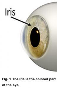
The colored part of the eye is called the iris. It is a circular muscle, similar in shape to a donut. The empty hole in the middle, which allows light to enter the eye, is called the pupil. When in a bright room or outdoors the pupil usually constricts; conversely when in a dark room the pupil usually dilates to allow more light to enter the eye [See figure 1].
Is it normal to have pupils of different sizes?
Normally the size of the pupil is the same in each eye, with both eyes dilating or constricting together. The term anisocoria refers to pupils that are different sizes at the same time. The presence of anisocoria can be normal (physiologic), or it can be a sign of an underlying medical condition.

When is anisocoria normal?
Approximately 20% of the population has anisocoria. The amount of anisocoria can vary from day-to-day and can even switch eyes. Anisocoria that is NOT associated with or due to an underlying medical condition is called physiologic anisocoria. Typically with physiologic anisocoria, the difference in pupil size between the two eyes does not exceed one millimeter.
How does the doctor determine whether anisocoria is due to an underlying medical problem?
Certain characteristics, such as when the anisocoria was first noted, whether it is more noticeable in bright or dim illumination, and whether or not there was a preceding event that could be related, will help determine the underlying cause. A complete eye examination by a pediatric ophthalmologist is performed and will evaluate the vision, eyelid position, how the eyes move, the health of the front and/or back portions of the eyes (among other things). The doctor will evaluate the size of the pupils and how they react to bright and dim lighting. Based on the evaluation, the doctor may wish to perform additional tests with eyedrops or perform laboratory or radiologic testing.
How does the doctor know if the big pupil is ‘too big’ or the small pupil is ‘too small’?
One of the most important parts in the evaluation of anisocoria is determining which pupil is abnormal. If the difference in size between the pupils increases in the dark, then the smaller (miotic) pupil may not be dilating well and could be the abnormal one. Conversely, if the difference in pupil size increases in bright lighting, then the larger (mydriatic) pupil may be the abnormal one because it is not constricting normally.
What are some causes of an abnormally large (dilated or mydriatic) pupil?
After trauma to the eye, the iris tissue can be injured causing the pupil to not constrict to bright light normally. Another possible cause is Adie’s tonic pupil syndrome. This is a condition most common in young adult females, which usually begins in one eye. The pupil is sluggish to react to light. Many people with this condition will also have diminished deep tendon reflexes and they can have trouble focusing at near. The condition is usually not associated with any more serious conditions. Some eyedrops have a dilating effect on the pupil, so eyedrop use is another cause of a dilated pupil. Finally, an abnormality of the third cranial nerve (a nerve that comes from the brain to the eye socket and controls eyelid position, eye movement, and pupil size) can cause a pupillary abnormality. In this condition, there is often ptosis (droopiness) of the upper eyelid on the same side as the dilated pupil. In addition, the eye may not move normally and an older child might complain of double vision. A third cranial nerve palsy can be a sign of a potentially serious condition, and the doctor may want to consider other possibilities.
What are some causes of an abnormally small (miotic) pupil?
Inflammation within the eye, whether from trauma or another cause, can result in a miotic pupil. Horner’s syndrome also produces a small pupil in the affected eye.
What are the signs of Horner’s syndrome?
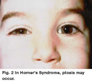
In Horner’s syndrome, the pupil in the involved eye is usually smaller and does not dilate as well as the other eye. The child may have mild ptosis (droopiness) of the upper eyelid [See figure 2]. A subtle but specific finding, which is sometimes present, is a slight elevation of the lower eyelid (known as inverse ptosis). Because the upper eyelid is slightly lower than normal, and the lower eyelid is slightly higher than normal, the eye may appear smaller. It is important to remember that if the droopy upper eyelid is on the side with the larger pupil, we are concerned about a pupil-involving third cranial nerve palsy and not Horner’s syndrome. A pupil-involving third cranial nerve palsy may be a sign of very serious medical conditions and requires immediate attention.
If the Horner’s syndrome developed during the first year of life, the iris on the affected side may appear lighter in color than the uninvolved side (heterochromia). Sometimes, the pressure in the eye is lower in the Horner’s eye and sometimes there is decreased sweating of the skin on the face on the affected side (anhydrosis).
What are the causes of Horner’s syndrome in children?

Horner’s syndrome can be divided into congenital (from birth) and acquired cases. Congenital Horner’s can result from neck trauma during birth and can be seen in association with a brachial plexus injury on the same side (Klumpke’s palsy). Often there is no apparent cause for congenital Horner’s. Acquired cases can be due to neck trauma, neck surgery, or an abnormality in the chest, neck or brain. In children, Horner’s syndrome may be caused by neuroblastoma, a tumor arising in another part of the body. Although rare, the risk of neuroblastoma is significantly greater with acquired Horner’s syndrome than it is with congenital cases.
What tests may be considered when Horner’s syndrome is suspected?
When clinical findings point towards a diagnosis of Horner’s syndrome, additional testing may be warranted. There are pharmacologic tests that the eye doctor may utilize to confirm a diagnosis of Horner’s syndrome, based on the response of the pupils to certain eye drops. When Horner’s syndrome is diagnosed in a child, the doctor may order additional tests including radiologic studies to help search for evidence of a neuroblastoma or other abnormalities in the abdomen, chest, or neck.
Credits: Journal of American Association for Pediatric Ophthalmology and Strabismus